

What are salivary Gland Stones (Sialolithasis)?
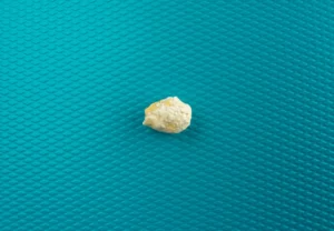
Salivary gland stones are also called sialoliths or salivary duct calculi, are mostly made up of inorganic matter. This matter commonly includes phosphate, calcium and magnesium. The stones vary widely in size and can measure up to 1.5cm in diameter.
There are hundreds of salivary glands around and in the mouth. Some of these glands are so small, you need a microscope to see them.
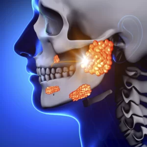 You have 3 sets of “major” salivary glands on each side. These are:
The parotid glands. These are located right in front of your ears and are your largest salivary glands.
The submandibular glands. These are under your jaw and are smaller. They produce saliva underneath your tongue.
The sublingual glands. These are the smallest and are located under the floor of your mouth below either side of your tongue.
Salivary stones can develop in any of your salivary glands or ducts. However, the submandibular gland and duct is more susceptible to developing stones than the other salivary glands.
The submandibular gland sends saliva to your mouth through a duct called Wharton’s duct, which has to run upward, fighting against gravity. This means the saliva does not always flow effectively and thus salivary gland stones can develop more easily. The saliva running through Wharton’s duct can also be a bit thicker than the saliva from your other glands, making it more prone to developing stones. Stones can also develop in the parotid gland and parotid duct, however this is less common as compared to the submandibular gland.
How do salivary gland stones (Sialolithasis) Occur?
 Salivary stones form when chemicals in the saliva accumulate in the duct or gland. However, the exact cause is not known.
Factors contributing to less saliva production and/or thickened saliva may be risk factors for salivary stones. These factors include: dehydration, poor eating, and use of certain medications including some antihistamines, blood pressure drugs, psychiatric drugs, and bladder control drugs. Trauma to the salivary glands may also raise the risk for salivary stones.
What are the symptoms of Sialolithasis?
Symptoms of sialolithasis may include: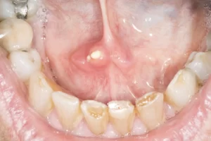
- A painful lump in the affected gland, with pain worsening during eating.
- A tender, painful lump in the cheek or under the chin.
- A foul-tasting discharge of pus from the duct into the mouth
- A lump or palpable stone on the floor of the mouth
In severe cases, fever, chills and general weakness may occur. There is also a possibility of developing a neck abscess (collection of pus in the neck) if the condition is severe.
How Is Sialolithasis Investigated?
Imaging
Imaging is frequently requested in order to diagnosis sialolithasis, the presence of a salivary gland stone and to assess for complications related to sialolithasis. This may take the form of ultrasound examinations, computed tomography (CT) scans, or Magnetic resonance imaging (MRI) scans.
Blood Tests
Routine blood tests are run to look for a high white blood count that would suggest a bacterial infection.
What are some medical treatments for sialolithasis?
Stay Hydrated
Hydration is extremely important if you have a salivary stone. On top of all the other benefits of water consumption, staying hydrated can increase your saliva production, which may help flush out the stone.
Apply a Warm Compress
Applying a warm compress such as a wet washcloth or a heating pad to the affected area could also help ease a little of the discomfort.
Encouraging saliva flow
Saliva flow can be increased by chewing sour, sugarless candies or by drinking orange or lemon juice.
What Are Some Surgical Treatments For Sialolithasis?
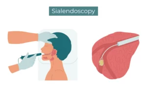 Sialendoscopy
Sialendoscopy is a procedure that is used to examine the ducts of the salivary glands. A miniature telescope, known as a endoscope, is inserted into the natural opening of the salivary gland duct as it enters the mouth. This allows the salivary gland ducts to be explored and small instruments can be inserted through the endoscope to remove any salivary stones or debris that may be present. The salivary glands can also be washed out with saline or other medications such as steroids and antibiotics.
Gland removal
Surgery to remove the entire salivary gland may be recommended in cases where sialendoscopy has failed or in cases of recurrent sialolithasis.